Virology tidbits

Virology tidbits

Wednesday 21 January 2015

Porcine Respiratory Syndrome Virus (PRRSV) and autophagy: a link between the ER stress response and p53?

Porcine reproductive and respiratory syndrome virus (PRRSV) is an enveloped virus with a positive strand ssRNA genome of approx. 15kB in length, encoding for ten open reading frames (ORFs).
Similar to the genome of the Coronaviridae, the PRRSV genome ORF1a and 1b genes encode for a RNA-dependent RNA Polymerase as well as for a number of non-structural proteins (nsp; 12 in total) which are generated by autoproteolytic cleavage by a virally encoded cysteine protease (nsp2), 3C-like serine protease (nsp4), and papain-like cysteine protease nsp1α/1β. As it is the case for the Coronaviridae, the remaining nsp’s of ORF1 encode for enzymes required for the replication of the viral genome, including a viral RNA Helicase (nsp-10) and endonuclease (nsp-11).
The remaining ORFs 2-7 encode for ORF2a (GP2a), ORF2b (E), ORF3 (GP3), ORF4 (GP4), ORF5a (GP5a), ORF5b (GP5b), ORF6 (M), and ORF7, the viral Nucleocapsid (N) which akin to the coronaviral N protein localises to the nucleolus and is phosphorylated.

PRRSV virus

PRRSV genome: ORFs and nsp's

In PRRSV infected MARC-145 cells, the viral nsp-2 protein is localised in the perinuclear region resembling a localisation at the ER, akin to the coronaviral proteases, thus suggesting that the expression of of nsp-2 may induce autophagy and a ER Stress response as described for the PLP2 and PLpro proteins derived from CoV. Indeed, PRRSV nsp-2 has been described to contain a DUB domain as well as inhibiting the Interferon response although an interaction with STING has not been demonstrated yet.  Although the data for the intracellular localisation of PRRSV structural proteins are incomplete, in the case of the related Equine Arterivirus (EAV), in BHK-21 cells the EAV E protein (encoded by ORF 2a) predominantly localises to the ER and to a lesser degree to the Golgi complex, whereas the viral GL  localises to the Golgi.

As described before for other positive strand RNA viruses such as Chikungunya Virus, Measles Virus, Coxsackie B Virus, Coronavirus as well as EMCV, the expression of viral proteins induces the formation of replication transcription centers (RTCs), double membrane  vesicles (DMV) which contain the viral RNA (both ssRNA and dsRNA intermediates) as well as the enzymes required for transcription including the viral RNA dependent RNA Polymerase and RNA Helicase. Commonly the DMV derive from the ER in a process subverting the autophagy pathway. As described before autophagy -which involves the formation of mature autophagosomes that fuse with lysosomes, ultimately leading to the degradation of the proteins localised within the autophagosome. Alternatively, the contents of autophagic vesicles might be secreted or in the case of viral proteins be processed to be displayed by MHC-Class I and MHC-Class II molecules.  As discussed before, viral proteins -sometimes the same which promote autophagy as for instance the CoV nsp-6 protein- not only promote the formation of DMV but also inhibit the formation of mature autophagosomes and/or the fusion of autophagosomes with lysosomes.

In the case of PRRSV, the viral nsp-2. nsp-3, and nsp-5/6/7 proteins have been demonstrated to localise to the ER and the expression of of the nsp-5/6/7 protein induces the formation of GFP-LC3 positive vesicles, indicating the induction of the formation of autophagosomes. Akin to the CoV nsp-6 protein, the expression of nsp-5/6/7 protein in Vero cells has been postulated to inhibit the fusion of the autophagosome with the lysosome. In the case of both nsp-2 and nsp-3 however the formation of autophagosomes has not been demonstrated (to my knowledge) although in MARC-145 cells and porcine pulmonary 
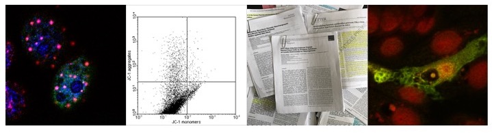
alveolar macrophages infected with PRRSV, LC3-II positive autophagosomes accumulate 24 hrs p.i. whilst the fusion with the lysosome is inhibited since the application of Chloroquine does not increase the number of GFP-RFP LC3-II positive punctae nor the levels of LC3-II as measured by western blot. Contrary to these results however, the treatment of PRRSV infected MARC-145 cells with Bafilomycin-A1 suggest that at 120 hrs p.i. PRRSV titers are decreased compared to mock treated cells and that the levels of p62/SQSTM-1 in PRRSV infected cells are lower than in non-infected cells. The difference observed might be due to the experimental conditions since Bafilomycin-A treatment lasted for 48 hrs compared to 6 hrs for Chloroquine treatment as well as different virus strains (PRRSV JXwn06 v. VR-2385), so more experiments are needed to address this issue.

Similar to the RTC induced following the infection of BHK-21 cells with EAV, these vesicles contain the viral nsp-2 and N protein although the presence of dsRNA has not been demonstrated to my knowledge. In contrast to EAV infected MEF, the autophagic machinery however is required for PRRSV replication  as viral titers are significantly lower in MARC-145 transfected with shLC3B, siATG7, siBeclin-1 or shATG5, suggesting that PRRSV -in contrast to EAV- does not induce the formation of autophagy-like vesicles via the ERAD pathway but via the induction of the phagophore via the ATG5/ATG7/Beclin-1 pathway; if however EDEMosomes are formed during PRRSV infection remains to be seen. Viral replication can also be induced by treating cells with Rapamycin, thus inhibiting mTORC1 and promoting autophagy, whereas treatment with 3-Methyladenine (3-MA) decreases viral titers.

Interestingly, the infection of MARC-145 with PRRSV strain VR-2385 activates mTORC1 (and thus inhibits autophagy) at early times post infection (6 h p.i.). So far the impact on viral or starvation induced autophagy has not been investigated, but the author these lines suggests that PRRSV inhibits autophagy at early timepoints p.i. whereas at later timepoints the formation of autophagy like vesicles is induced. This hypothesis is supported by results indicating that PIK-K-Akt kinase signalling is modulate by PRRSV in so far as phosphorylated Akt kinase levels increase at earlier timepoints, but decrease at 12 hrs p.i.  . It is however crucial to compare proteins derived from highly virulent strains to those derived from attenuated or less pathogenic strains. 

Induction of p53 and DRAM-1 dependent autophagy via the ER stress by
PRRSV: hypothetical model

PRRSV and the ER stress response: does nsp-2, nsp-4, or nsp-5/6/7 induce the
ER stress response?

It remains therefore to be seen if the expression of PRRSV proteins increases the formation of autophagosomes and/or autophagy-like vesicles similar to the coronaviral nsp-3/-4/-6 proteins whilst inhibiting the fusion of the lysosome. Also, it remains to be seen if the expression of PRRSV nsp-2 -and other viral proteins including nsp-5/6/7 induces the ER stress response by lipid depletion and subsequent autosis. Interestingly the infection of MARC-145 cells with PRRSV strain CH-1a results in a PERK and IRE1 induced ER stress response whose inhibition is associated with decreased viral replication. Since autophagy is induced fooling the activation of the ER stress response it seems possible that the decrease in viral replication is due to a decrease in autophagy or alternatively to apoptosis (autophagy dependent or independent). In this case, the activation of the ER stress response might induce p53 and thus DRAM-1; indeed, the inhibition of p53 has been demonstrated to decrease viral titers, but so far no link has been established between PRRSV, the ER stress response, p53, and autophagy.  


Interplay of the induction of Akt, Akt dependent inhibition of autophagy and induction
of autophagy via the ER stress response: activation of Akt early during the infection, induction of the ER
stress response late in infection?


ResearchBlogging.org


Further reading

Meulenberg, J. (2000). PRRSV, the virus Veterinary Research, 31 (1), 11-21 DOI: 10.1051/vetres:2000103 

Sun Z, Chen Z, Lawson SR, & Fang Y (2010). The cysteine protease domain of porcine reproductive and respiratory syndrome virus nonstructural protein 2 possesses deubiquitinating and interferon antagonism functions. Journal of virology, 84 (15), 7832-46 PMID: 20504922 

Shi X, Zhang G, Wang L, Li X, Zhi Y, Wang F, Fan J, & Deng R (2011). The nonstructural protein 1 papain-like cysteine protease was necessary for porcine reproductive and respiratory syndrome virus nonstructural protein 1 to inhibit interferon-β induction. DNA and cell biology, 30 (6), 355-62 PMID: 21438756 

You JH, Howell G, Pattnaik AK, Osorio FA, & Hiscox JA (2008). A model for the dynamic nuclear/nucleolar/cytoplasmic trafficking of the porcine reproductive and respiratory syndrome virus (PRRSV) nucleocapsid protein based on live cell imaging. Virology, 378 (1), 34-47 PMID: 18550142

Chen Z, Zhou X, Lunney JK, Lawson S, Sun Z, Brown E, Christopher-Hennings J, Knudsen D, Nelson E, & Fang Y (2010). Immunodominant epitopes in nsp2 of porcine reproductive and respiratory syndrome virus are dispensable for replication, but play an important role in modulation of the host immune response. The Journal of general virology, 91 (Pt 4), 1047-57 PMID: 19923257

Fang Y, Treffers EE, Li Y, Tas A, Sun Z, van der Meer Y, de Ru AH, van Veelen PA, Atkins JF, Snijder EJ, & Firth AE (2012). Efficient -2 frameshifting by mammalian ribosomes to synthesize an additional arterivirus protein. Proceedings of the National Academy of Sciences of the United States of America, 109 (43) PMID: 23043113 

Snijder EJ, van Tol H, Pedersen KW, Raamsman MJ, & de Vries AA (1999). Identification of a novel structural protein of arteriviruses. Journal of virology, 73 (8), 6335-45 PMID: 10400725 

Oleksiewicz MB, & Nielsen J (1999). Effect of porcine reproductive and respiratory syndrome virus (PRRSV) on alveolar lung macrophage survival and function. Veterinary microbiology, 66 (1), 15-27 PMID: 10223319 

 Monastyrska I, Ulasli M, Rottier PJ, Guan JL, Reggiori F, & de Haan CA (2013). An autophagy-independent role for LC3 in equine arteritis virus replication. Autophagy, 9 (2), 164-74 PMID: 23182945 

Knoops K, Bárcena M, Limpens RW, Koster AJ, Mommaas AM, & Snijder EJ (2012). Ultrastructural characterization of arterivirus replication structures: reshaping the endoplasmic reticulum to accommodate viral RNA synthesis. Journal of virology, 86 (5), 2474-87 PMID: 22190716 

Lu W, Sun B, Mo J, Zeng X, Zhang G, Wang L, Zhou Q, Zhu L, Li Z, Xie Q, Bi Y, & Ma J (2014). Attenuation and immunogenicity of a live high pathogenic PRRSV vaccine candidate with a 32-amino acid deletion in the nsp2 protein. Journal of immunology research, 2014 PMID: 25009824

Pujhari S, Kryworuchko M, & Zakhartchouk AN (2014). Role of phosphatidylinositol-3-kinase (PI3K) and the mammalian target of rapamycin (mTOR) signalling pathways in porcine reproductive and respiratory syndrome virus (PRRSV) replication. Virus research, 194, 138-44 PMID: 25304692

Cottam EM, Whelband MC, & Wileman T (2014). Coronavirus NSP6 restricts autophagosome expansion. Autophagy, 10 (8), 1426-41 PMID: 24991833 S

Sun MX, Huang L, Wang R, Yu YL, Li C, Li PP, Hu XC, Hao HP, Ishag HA, & Mao X (2012). Porcine reproductive and respiratory syndrome virus induces autophagy to promote virus replication. Autophagy, 8 (10), 1434-47 PMID: 22739997 

Liu Q, Qin Y, Zhou L, Kou Q, Guo X, Ge X, Yang H, & Hu H (2012). Autophagy sustains the replication of porcine reproductive and respiratory virus in host cells. Virology, 429 (2), 136-47 PMID: 22564420 

Chen Q, Fang L, Wang D, Wang S, Li P, Li M, Luo R, Chen H, & Xiao S (2012). Induction of autophagy enhances porcine reproductive and respiratory syndrome virus replication. Virus research, 163 (2), 650-5 PMID: 22119900 

Cottam EM, Maier HJ, Manifava M, Vaux LC, Chandra-Schoenfelder P, Gerner W, Britton P, Ktistakis NT, & Wileman T (2011). Coronavirus nsp6 proteins generate autophagosomes from the endoplasmic reticulum via an omegasome intermediate. Autophagy, 7 (11), 1335-47 PMID: 21799305 

Huo Y, Fan L, Yin S, Dong Y, Guo X, Yang H, & Hu H (2013). Involvement of unfolded protein response, p53 and Akt in modulation of porcine reproductive and respiratory syndrome virus-mediated JNK activation. Virology, 444 (1-2), 233-40 PMID: 23850458

Muñoz-Pinedo, C., & Martin, S. (2014). Autosis: a new addition to the cell death tower of babel Cell Death and Disease, 5 (7) DOI: 10.1038/cddis.2014.246 

Liu Y, Shoji-Kawata S, Sumpter RM Jr, Wei Y, Ginet V, Zhang L, Posner B, Tran KA, Green DR, Xavier RJ, Shaw SY, Clarke PG, Puyal J, & Levine B (2013). Autosis is a Na+,K+-ATPase-regulated form of cell death triggered by autophagy-inducing peptides, starvation, and hypoxia-ischemia. Proceedings of the National Academy of Sciences of the United States of America, 110 (51), 20364-71 PMID: 24277826

Thursday 15 January 2015

KSHV and autophagy: vFLIP, v-CyclinD, and v-Bcl2

As discussed in various posts before, viral proteins interfere with autophagy at various points to promote viral replication either by subverting the cellular autophagy machinery to form replication centers, to prevent the presentation of viral antigen via the MHC Class -I/-II complex, interfere with antiviral signalling or to prevent the degradation of viral proteins and thus increase viral replication.  The localisation of viral proteins to the ER can induce ER stress and thus promote autophagy, thus counteracting or delaying apoptosis, whereas other viral proteins derived from the same virus inhibit the fusion with the lysosome or prevent the maturation of the autophagosome by various mechanisms including binding to Beclin-1.  Deregulation of autophagy however is not limited to cells infected with viruses or bacteria but can also be observed in human cancers. In the context of infection of cells with oncogenic viruses, interference with the autophagy pathway has been documented for proteins derived from a variety of oncogenic viruses, including but not limited to proteins encoded by Human T-Cell Leukemia/Lymphoma Virus-1 (HTLV-1), JC virus (JCV), BK polyomavirus (BKPyV), Epstein-Barr virus (EBV), Kaposi’s Sarcoma-Associated Herpesvirus (KSHV), Hepatitis B virus (HBV), and Hepatitis C virus (HCV). Akin to the infection of cells with positive strand RNA viruses, the induction of autophagy can have positive effects (e.g. BKPyV) or negative effects (e.g. JCV) on viral replication. Since the expression of viral oncoprotein commonly also induces the DNA damage response and thus might promote autophagy via increasing the expression of pro-autophagy genes in a p53-dependent manner as discussed before, oncogenic viruses the regulation of autophagy might contribute to the transformation of cells both in vitro and in vivo.

Kaposi Sarcoma- Associated Herpesvirus (KSHV) - also known as Human Herpesvirus 8 (HHV8) - is the causative agent of Kaposi’s Sarcoma (KS), a cancer associated with prominent cutaneous lesions. Being a Herpesvirus, KSHV is a large dsDNA virus with genome of approx. 165 kb in size. An amorphous protein layer, the tegument, surrounds the capsid and a lipid envelope derived from the cellular plasma membrane. The genome itself encodes for a number of viral variants of cellular proteins including Cyclin D (v-Cyclin D), Flice inhibitory protein (v-FLIP), and Bcl-2 (v-Bcl2) as well as Latency-associated nuclear antigen (LANA) which has a p53 binding activity that are required for the transformation of primary spindle cells infected with KSHV into tumour cells.

                                  v-Cyclin D and v-Bcl2

v-Cyclin D is a homologue of the cellular Cyclin D and as such forms an active holoenzyme with both Cyclin dependent kinases  (CDK) -4 and -6. Unlike cellular Cyclin D however, v-Cyclin D also associates with CDK-2 and CDK-9 in addition to having a broader substrate range than cellular Cyclin D. Complexes of v-Cyclin D and CDK-6 are also refractory to Cip/Kip and INK4 families of Cyclin-dependent kinase inhibitors (CKIs). Consequently, v-Cyclin D has been demonstrated to increase DNA synthesis via phosphorylation of Orc1 and Cdc6 in tumour cell lines whilst inducing the DNA damage response via activation of the ATM-Chk2 pathway in hTERT immortalised and primary human dermal microvascular endothelial cells  (HDMEC) as evidenced by increase in γH2AX and 53BP1 positive foci. Similar to HDMEC expressing H-RasV12 , the expression of induces either autophagy dependent apoptosis or an irreversible cell cycle arrest known as oncogene-induced senescence (OIS).
As discussed before the activation of p53 by various pathways, including ATM and ATR, induces the formation of the autophagosome by increasing the expression of several genes, including DRAM and Sestrin-1. In the case of KSHV v-Cyclin D, in TIME cells expressing v-Cyclin D, increased levels of DRAM and Sestrin-1 have been observed as well as increased levels of p53 phosphorylated at Ser-15, indicating that the expression of KSHV v-Cyclin D induces autophagy via activation of p53 by ATM but not ATR. Interestingly, the expression of LANA does not counteract v-Cyclin D induced autophagy, reflecting experimental data that only a small amount of LANA binds p53.


KSHV v-Cyclin D and autophagy: induction of OIS via DRAM-1 ?


Since the induction of autophagy by H-RasV12 has been linked to the induction of OIS, the expression of v-Cyclin D may also induce OIS by a similar mechanism. KSHV infected cells however do not display OIS, suggesting that the viral genome encodes proteins inhibiting autophagy. One of these proteins is v-Bcl2 which similar to cellular Bcl-2 binds Beclin-1 and thus prevents the formation of the phagophore. In the case of v-Bcl2 derived from the murine γ-Herpesvirus 68 (γHV68), a mutant variant of γHV68 which expresses a v-Bcl2 variant that does not bind Beclin-1 does not maintain a chronic infection in mice whereas a mutant unable to bind the pro-apoptotic Bak protein establishes chronic infections   comparable to wt virus, indicating that the ability to inhibit autophagy is required to establish a chronic infection. In the case of KSHV v-Bcl2, cellular assays indicate that v-Bcl2 does not bind Bak but in vitro studies using purified proteins suggest that v-Bcl2 can bind Bak. 

Inhibition of v-Cyclin D induced autophagy by v-Bcl2 and vFLIP prevents OIS

The importance of inhibiting autophagy in KSHV infected cells is further highlighted by data indicating that in HEK 293T cells infected with KSHV the viral replication and transcription activator (RTA) encoded by KSHV ORF 50 increases the expression of cellular Bcl-2 following KSHV reactivation; indeed in cells transfected with shRNA targeting cellular Bcl-2, increased apoptosis and decreased viral replication can be observed (if the formation of autophagosomes is decreased as well has not been investigated).If similar to EBV RTA the expression of other autophagy related genes is also induced remains to be seen.  


                                           v-FLIP

The viral Fas-associated death domain-like interleukin-1β (IL-1β)-converting enzyme-inhibitory protein (vFLIP) has been postulated to bind to caspase-8 -and thus prevent caspase-8/Fas mediated apoptosis- although the initial results could not be confirmed in transgenic mice expressing vFLIP. Instead, vFLIP has been shown to regulatory components of the IκB kinase signalosome (in particular NEMO and IKKαβγ), inducing the expression of anti-apoptotic proteins such as cellular inhibitor of apoptosis (cIAP)-1 and -2 as well as c-FLIP via NF-κB dependent signaling pathways inducing the expression of cytokines. Unlike cFLIP, binding of vFLIP to NEMO is independent of TRAF thus constitutively activating NF-κB signalling pathways.


Inducing autophagy of a lymphoma cell line positive for KSHV, BCBL-1, and HEK 293T cells expressing a KSHV-ΔvFLIP construct by Rapamycin treatment induces growth arrest and autophagy-induced apoptosis, which can be attenuated by transfection of Beclin-1 siRNA, indicating that the induction of autophagy by KSHV is indeed sufficient to induce apoptosis. Autophagy induced cell death however can also be prevented by transfection of plasmids allowing the expression of  wt vFLIP or mutant vFLIP carrying a deletion of the Fas-Associated protein with Death Domain (FADD), indicating that the ability of vFLIP to bind caspase-8 is not required for preventing apoptosis. Human NIH 3T3 and HCT 116 cells or murine MEF expressing vFLIP derived from KSHV, Herpesvirus Saimiri (HSV), or  Molluscum contagiosum virus (MCV)  were able to prevent starvation induced autophagy and NIH 3T3 cells expressing an inducible Trex-BCBL-vFLIP plasmid reduced the formation of autophagosomes (as well as  growth arrest and apoptosis) compared to cells expressing the Trex-BCBL plasmid following Rapamycin treatment, indicating that vFLIP indeed is sufficient to inhibit autophagy. When immunoprecipated, KSHV vFLIP was shown to bind Atg3 via an Atg3 binding domain, thus preventing the conversion of LC3-I to LC3-II; accordingly, the expression of vFLIP whilst not preventing the formation of LC3-I positive autophagic vesicles inhibits the formation of LC3-II positive autophagosomes. Taken together with v-Bcl2 however the expression of both proteins together should prevent the formation of autophagy-related vesicles.
Since the expression of KSHV v-Cyclin D induces autophagy -and thus OIS- as discussed above, the expression of both v-Bcl2 and vFLIP counteracts OIS, allowing increased cell proliferation. It should be noted however that preventing OIS also leads to a mitotic catastrophe and aberrant centrosome duplication, both which are common features of tumour cells and indeed can be observed in KSHV infected cells. Inhibiting autophagy however also inhibits the secretion of cytokines as well as preventing the expression of viral latent proteins. It should be noted that the ectopic expression of a DED1 alpha2-helix ten amino-acid (alpha2) peptide or a DED2 alpha4-helix twelve amino-acid (alpha4) peptide containing the vFLIP-Atg3 binding domain in KSHV infected cells induces autophagy and autophagy associated apoptosis probably by sequestering vFLIP, preventing the interaction of vFLIP with Atg3.

vFLIP and autophagy:inhibition via binding to ATG3


Recent data indicate that the inhibition of autophagy KSHV vFLIP in endothelial Long-Term-Infected Telomerase-Immortalized Endothelial Cells (TIVE-LTC), leads to an accumulation of Ser-403 phosphorylated p62/SQSTM-1 in the cytoplasm. As discussed before, Ser-403 phosphorylated p62/SQSTM-1 can induce the ubquitinylation of Keap1 and its degradation via the proteasome concomitant with stabilisation and clear translocation of Nrf2. Indeed, vFLIP induces the nuclear translation of Nrf2 in TIVE-LTC cells as well as induction of genes related to angiogenesis, inflammation, and the antioxidant response. In TIVE-LTC the stabilisation of Nrf2 is increased via vFLIP mediated activation of PKCζ via cycloxygenase-2 (COX-2)/PGE-2 mediated signalling pathways. It remains to be seen however if the expression of the vFLIP-Atg3 binding domain can reverse the increase of Nrf2 by stimulating p62/SQSTM-1 dependent selective autophagy.


KSHV vFLIP and degradation of Keap1 via increased levels of p62/SQSTM-1 and
PKCζ

v-Cyclin D, v-Bcl2, vFLIP are however not the only KSHV proteins interfering with autophagy. Proteins which induce the formation of the phagophore include the K1, vIL-6, and a viral G protein­ coupled receptor (vGPCR) of KSHV, all of which activate the Phosphoinositide 3­kinase (PI3K)– AKT–mTORC1 pathway and the ORF45 protein has been shown to contribute to the activation of mTORC1 in lymphatic endothelial cells by activating the ERK2–RSK1–TSC2 pathway. KSHV K7 inhibits the maturation of autophagosomes by binding Rubicon thus preventing the fusion of the autophagosome with the lysosome. 

The expression of K7,  vBcl-2 and vFLIP therefore is central to the ability of KSHV to evade antiviral signalling pathways whilst maintaining signalling required for cellular survival and proliferation. 
Another viral protein, ORF 57, has been shown to increase the expression of X-linked inhibitor of apoptosis (XIAP). Interestingly, XIAP has been linked to autophagy inhibition via degradation of Mdm2 and stabilization of p53 in serum starved MEF, which has been linked to increased tumourgenesis in mice. The question remains if this is also true for cells –murine or human- expressing KSHV or the murine γHV68. Interestingly, the expression of Rhesus monkey rhadinovirus (RRV) derived vFLIP in Hela  as well as in RRV infected BJAB cells inhibits apoptosis whilst increasing the formation of LC3-II positive autophagosomes. So far however a smilier effect has not been reported in cells derived from rabbits.  


KSHV and autophagy: summary 

Recent studies involving drugs such as Sirolimus which activate autophagy by binding mTOR and thus inhibiting the mTOR kinase highlighted the importance of autophagy inhibition in KSHV infected cells, delaying IL-6, IL-10, and IFN-γ dependent tumourgenesis.


ResearchBlogging.org


Further reading


Silva LM, & Jung JU (2013). Modulation of the autophagy pathway by human tumor viruses. Seminars in cancer biology, 23 (5), 323-8 PMID: 23727156 


Sariyer IK, Merabova N, Patel PK, Knezevic T, Rosati A, Turco MC, & Khalili K (2012). Bag3-induced autophagy is associated with degradation of JCV oncoprotein, T-Ag. PloS one, 7 (9) PMID: 22984599

Bouley SJ, Maginnis MS, Derdowski A, Gee GV, O'Hara BA, Nelson CD, Bara AM, Atwood WJ, & Dugan AS (2014). Host cell autophagy promotes BK virus infection. Virology, 456-457, 87-95 PMID: 24889228

Leidal AM, Cyr DP, Hill RJ, Lee PW, & McCormick C (2012). Subversion of autophagy by Kaposi's sarcoma-associated herpesvirus impairs oncogene-induced senescence. Cell host & microbe, 11 (2), 167-80 PMID: 22341465

Pratt ZL, & Sugden B (2012). How human tumor viruses make use of autophagy. Cells, 1 (3), 617-30 PMID: 24710493

Pekkonen P, Järviluoma A, Zinovkina N, Cvrljevic A, Prakash S, Westermarck J, Evan GI, Cesarman E, Verschuren EW, & Ojala PM (2014). KSHV viral cyclin interferes with T-cell development and induces lymphoma through Cdk6 and Notch activation in vivo. Cell cycle (Georgetown, Tex.), 13 (23), 3670-84 PMID: 25483078

Zhi H, Zahoor MA, Shudofsky AM, & Giam CZ (2014). KSHV vCyclin counters the senescence/G1 arrest response triggered by NF-κB hyperactivation. Oncogene PMID: 24469036

E X, Hwang S, Oh S, Lee JS, Jeong JH, Gwack Y, Kowalik TF, Sun R, Jung JU, & Liang C (2009). Viral Bcl-2-mediated evasion of autophagy aids chronic infection of gammaherpesvirus 68. PLoS pathogens, 5 (10) PMID: 19816569


Münz C (2011). Beclin-1 targeting for viral immune escape. Viruses, 3 (7), 1166-78 PMID: 21994775 Wirawan E, Lippens S, Vanden Berghe T, Romagnoli A, Fimia GM, Piacentini M, & Vandenabeele P (2012). Beclin1: a role in membrane dynamics and beyond. Autophagy, 8 (1), 6-17 PMID: 22170155


Su M, Mei Y, Sanishvili R, Levine B, Colbert CL, & Sinha S (2014). Targeting γ-herpesvirus 68 Bcl-2-mediated down-regulation of autophagy. The Journal of biological chemistry, 289 (12), 8029-40 PMID: 24443581


Wen HJ, Yang Z, Zhou Y, & Wood C (2010). Enhancement of autophagy during lytic replication by the Kaposi's sarcoma-associated herpesvirus replication and transcription activator. Journal of virology, 84 (15), 7448-58 PMID: 20484505


Santarelli R, Gonnella R, Di Giovenale G, Cuomo L, Capobianchi A, Granato M, Gentile G, Faggioni A, & Cirone M (2014). STAT3 activation by KSHV correlates with IL-10, IL-6 and IL-23 release and an autophagic block in dendritic cells. Scientific reports, 4 PMID: 24577500

Hung CH, Chen LW, Wang WH, Chang PJ, Chiu YF, Hung CC, Lin YJ, Liou JY, Tsai WJ, Hung CL, & Liu ST (2014). Regulation of autophagic activation by Rta of Epstein-Barr virus via the extracellular signal-regulated kinase pathway. Journal of virology, 88 (20), 12133-45 PMID: 25122800 

Kalt I, Borodianskiy-Shteinberg T, Schachor A, & Sarid R (2010). GLTSCR2/PICT-1, a putative tumor suppressor gene product, induces the nucleolar targeting of the Kaposi's sarcoma-associated herpesvirus KS-Bcl-2 protein. Journal of virology, 84 (6), 2935-45 PMID: 20042497

Ritthipichai K, Nan Y, Bossis I, & Zhang Y (2012). Viral FLICE inhibitory protein of rhesus monkey rhadinovirus inhibits apoptosis by enhancing autophagosome formation. PloS one, 7 (6) PMID: 22745754

Lee JS, Li Q, Lee JY, Lee SH, Jeong JH, Lee HR, Chang H, Zhou FC, Gao SJ, Liang C, & Jung JU (2009). FLIP-mediated autophagy regulation in cell death control. Nature cell biology, 11 (11), 1355-62 PMID: 19838173

Gjyshi O, Flaherty S, Veettil MV, Johnson KE, Chandran B, & Bottero V (2014). Kaposi's Sarcoma-Associated Herpesvirus Induces Nrf2 Activation in Latently-Infected Endothelial Cells Through SQSTM1 Phosphorylation and Interaction with Polyubiquitinated Keap1. Journal of virology PMID: 25505069

Liang Q, Chang B, Brulois KF, Castro K, Min CK, Rodgers MA, Shi M, Ge J, Feng P, Oh BH, & Jung JU (2013). Kaposi's sarcoma-associated herpesvirus K7 modulates Rubicon-mediated inhibition of autophagosome maturation. Journal of virology, 87 (22), 12499-503 PMID: 24027317


Liang C, Oh BH, & Jung JU (2015). Novel functions of viral anti-apoptotic factors. Nature reviews. Microbiology, 13 (1), 7-12 PMID: 25363821

Nishimura K, Ueda K, Guwanan E, Sakakibara S, Do E, Osaki E, Yada K, Okuno T, & Yamanishi K (2004). A posttranscriptional regulator of Kaposi's sarcoma-associated herpesvirus interacts with RNA-binding protein PCBP1 and controls gene expression through the IRES. Virology, 325 (2), 364-78 PMID: 15246275

Huang X, Wu Z, Mei Y, & Wu M (2013). XIAP inhibits autophagy via XIAP-Mdm2-p53 signalling. The EMBO journal, 32 (16), 2204-16 PMID: 23749209

Lambert PJ, Shahrier AZ, Whitman AG, Dyson OF, Reber AJ, McCubrey JA, & Akula SM (2007). Targeting the PI3K and MAPK pathways to treat Kaposi's-sarcoma-associated herpes virus infection and pathogenesis. Expert opinion on therapeutic targets, 11 (5), 589-99 PMID: 17465719